On March 5, the research team led by Professors YU Yanqin, DUAN Shumin and YANG Hongbin from the Zhejiang University School of Brain Science and Brain Medicine published an open-access article titled “Control of defensive behavior by the nucleus of Darkschewitsch GABAergic neurons” in the journal National Science Review. This groundbreaking work reveals, for the first time, the crucial role of GABAergic neurons in the nucleus of Darkschewitsch (ND) in modulating defensive behavior, thereby expanding our understanding of the neural mechanisms underlying fear defense.
In recent decades, significant strides have been made in identifying the roles of the amygdala, the hypothalamus, and the periaqueductal gray in defensive behavior. Researchers have elucidated specific circuits governing defensive responses to various threat stimuli. However, due to the complexity of brain circuits, many nuclei and circuits involved in the regulation of defensive responses remain to be explored. Moreover, there remains scanty research into brain regions that can broadly regulate defensive responses to various types of threats, and the mechanisms by which they specifically regulate a particular defensive behavior are still elusive.
The ND, named after Russian neurologist L. O. Darkshevich, was first described in 1889 as an accessory oculomotor nucleus and was postulated in controlling eye movement via zone second cervical spinal cord of the flocculus. However, there has long been a paucity of research on the anatomical outputs and inputs and the physiological functions of GABAergic neurons of the ND.
In this work, the researchers selected two different types of fear stimuli: odorant trimethylthiazoline (TMT) and foot shocks. When mice were exposed to these stimuli, the NDGABA neurons were significantly activated. Further in situ fiber photometry revealed a substantial rise in calcium (Ca2+) activity in NDGABA neurons. Notably, through electrophysiological recordings, the researchers observed that NDGABA neurons specifically influenced freezing-like behavioral responses, pointing to the relationship between NDGABA neurons and freezing-like defensive behavior.
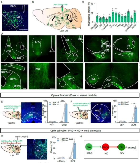
To investigate the causal relationship between NDGABA neurons and defensive behavior, the researchers employed optogenetics to activate NDGABA neurons, and found that it immediately induced freezing-like behavior in mice rather than escape or hiding responses. It also brought about changes in autonomic functions associated with freezing behavior, such as an increased pupil size and a decreased heart rate, and aversion. Conversely, optogenetic inhibition of NDGABA neurons significantly reduced freezing behavior in both innate and conditioned fear, without impacting locomotion and avoidance behavior, indicating the pivotal role of NDGABA neurons in freezing-like behavior. Through a combination of retrograde and anterograde viral tracing alongside optogenetic techniques, the researchers further identified that NDGABA neurons receive inputs from glutamatergic neurons in the lateral periaqueductal gray (lPAG). The ND is responsible for transmitting fear information from the lPAG to glutamatergic neurons in the ventral part of the gigantocellular nucleus (GiV), ultimately triggering freezing behavior. Interestingly, GABAergic neurons expressing somatostatin (SOM), rather than parvalbumin (PV), were found to be crucial in controlling freezing-like behavior.
This study sheds light on the specific control of freezing defensive responses by the ND brain region and the lPAGglu-NDGABA-GiVglu circuit, deepening our understanding of the functionality of the ND brain region. It supplements our existing knowledge on the downstream mechanisms of fear control by the PAG and the circuitry network map of the “fear system” in the brain, offering fresh insights into the biological basis of fear defensive behavior. Additionally, given the regional conservatism of the ND brain region in rodents and humans, this study may present a promising target for treating fear disorders and other psychiatric illnesses.

NDGABA neurons relay fearful information from PAG to the medulla